UNDERSTANDING YOUR EXAMINATION
What is an
MRI Scan?
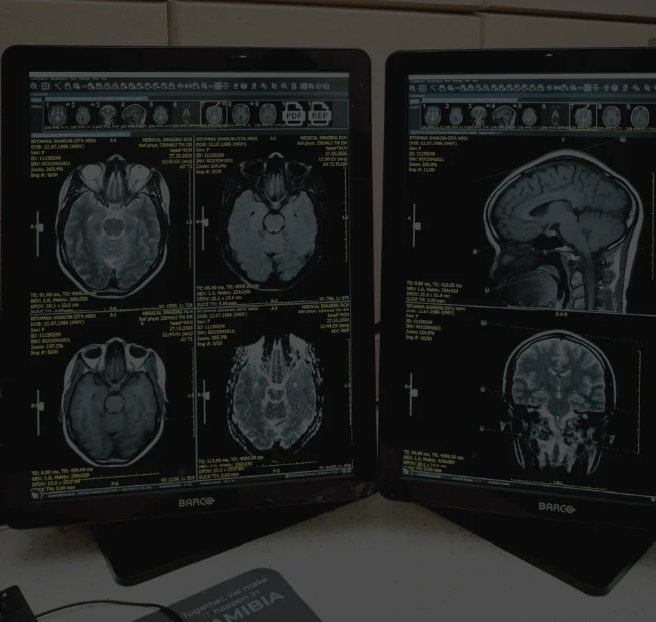
An MRI scan uses a strong magnetic field and radio waves to produce detailed images of organs, soft tissues, bones, and other structures inside your body. Working together, these advanced technologies allow clear, cross-sectional views without using ionising radiation.
These images help our radiologists assess the brain, spine, joints, and internal organs, giving your referring doctor valuable information to guide your care.
DETAILED SOFT TISSUE IMAGING
Clear images help radiologists examine the brain, spine, muscles, ligaments, and internal organs.
RADIATION-FREE TECHNOLOGY
Uses magnetic fields and radio waves to capture diagnostic images without ionising radiation.
COMPREHENSIVE DIAGNOSTIC INSIGHTS
Supports a wide range of evaluations, including neurological, musculoskeletal, and vascular assessments.
SPECIALIST INTERPRETATION
Experienced radiologists review your images to help guide your referring doctor’s decisions.